
Anatomical literature and drawings – digitized
 In Cooperation with the Institute for Anatomy and Cell Biology, the Library of the University of Heidelberg has digitalized chosen anatomical literature and lithographs from the 19th century. This includes textbooks, scripts and drawings which described the teaching and research of this period, and has provided a unique insight into the work and history of the Institute. But historical collection journals can also be viewed in digital form. They provide a good indication of the teaching priorities of the respective chair holders, or of the research interests they pursued. Their importance is growing, especially in the context of provenance research, as they represent a unique source for the time when the preparations and models were created.
Search
Start browsing by:
The oldest textbooks that have been digitalized were written by Jacob Fidelis Ackermann (*1765 †1815). The public was made aware of his presence from the beginning of his tenure in Heidelberg because he attempted, often with little success, to rebut the phrenological theses of Franz Josef Galls (*1758 †1828). Ackermann also actively practiced medicine. In addition he founded an outpatient clinic, in which he volunteered his time to treat the poor. This earned him the respect and admiration of the people.
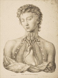
Most of the anatomical drawings are lithographs by anatomists who worked in Heidelberg during their scientific career. Many of these very artistic lithographs originated in Heidelberg. Indeed Friedrich Tiedemann (*1781 †1861) dissected innumerable human and zoological cadavers during his tenure in Heidelberg, which he used to write his book “Tabulae Anatomicae”. Unfortunately, the work of Vincent Fohmann (*1794 †1837) concerning the “Saugadersystem”, today known as the lymphatic system, has been for all practical purpsoes forgotten. He was however well known outside of Germany for his uncomparable detailed mercury injections of specimens. The collected works of Jacob Henle (*1809 †1885) provide information conserning the Institute’s extensive collection. He provided detailed information about the anatomical and pathological dissections, that were produced around 1844 in the Heidelberg collection. Henle and Tiedemann then as now were very popular anatomists.
Other authors include the Anatomists Friedrich Arnold (*1803 †1890), Carl Gegenbaur (*1826 †1903), and Alexander Ecker (*1816 †1887). Arnold is still well known today for his research on the human brain. His plates primarily displayed the brain, spinal cord, sensory organs as well as bones. Gegenbaur compared the structure of humans with that of animals (“Vergleichende Anatomie”), in 1869 Ecker suggested the nomenclature for the human gyri and sulci, terms that are still used today. In addition he was interested in anthropology.
- Jacob Fidelis Ackermann
- Friedrich Arnold
- Theodor Ludwig Wilhelm Bischoff
- Hermann Braus
- Carl Wilhelm Ludwig Bruch
- Vincent Fohmann
- Max Fürbringer
- Alexander Ecker
- Carl Gegenbaur
- Jacob Henle
- Georg Ludwig Kobelt
- Johann Anton Nuhn
- Friedrich Tiedemann
- August Vierling
In addition to the historical publications, hundreds of plates, pictures, photographs and illustrations can be accessed through the internet portal heidICON. The large formated plates were produced in the beginning of the 20th century and presented during the medical lectures. Students were given the opportunity to borrow the photographs and illustrations together with the models and dissections.
Requests, suggestions and criticism to Dr.sc.hum. Sara Doll
Further information
Historical Anatomies on the Web (U.S. National Library of Medicine)
 Universitaetsbibliothek Heidelberg